
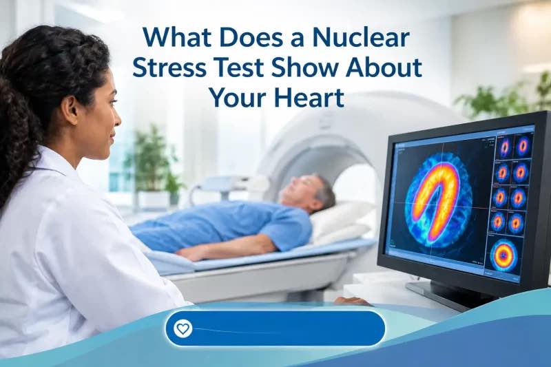
Chest discomfort, shortness of breath, unusual fatigue, or an abnormal ECG can leave patients with an unsettling question: Is the heart getting enough blood when it has to work harder?
That is one reason doctors may order a nuclear stress test.
So, what does a nuclear stress test show? It provides images of blood flow to the heart muscle at rest and during stress. The test can help doctors identify areas that may not be receiving enough blood, assess possible coronary artery disease, look for evidence of previous heart damage, and evaluate how well the heart is pumping.
A nuclear stress test is also called myocardial perfusion imaging (MPI). It uses a small amount of radioactive tracer and a specialized camera—usually SPECT or PET—to create images of blood flow through the heart. The American Heart Association explains how myocardial perfusion imaging works, including how PET and SPECT scans are used to evaluate blood flow and heart function.
Despite the word “nuclear,” this is a commonly used diagnostic imaging test, not radiation treatment.
What Does a Nuclear Stress Test Measure?
A nuclear stress test combines information about blood flow to the heart muscle with information about how the heart responds to physical or medication-induced stress.
Patients generally have images taken while the heart is at rest and again after the heart has been stressed. Stress may come from walking on a treadmill or, for people who cannot exercise adequately, from medication that changes blood flow or otherwise stresses the cardiovascular system.
The radioactive tracer travels through the bloodstream and is taken up by heart muscle. Areas receiving adequate blood flow generally show tracer uptake on the images. Areas with reduced uptake may indicate reduced blood flow, previous heart damage, or sometimes an imaging artifact. RadiologyInfo’s overview of cardiac nuclear medicine explains how the tracer and imaging equipment are used to evaluate blood flow and heart function.
A nuclear stress test may show:
- Normal blood flow at rest and during stress. This generally suggests that the scan did not identify a significant stress-related perfusion abnormality.
- Reduced blood flow during stress but not at rest. This is often described as a reversible perfusion defect and may suggest ischemia—meaning the heart muscle is not receiving enough blood when demand increases.
- Reduced blood flow at rest and during stress. This is sometimes called a fixed defect. It can reflect previous heart muscle damage or scar, although artifacts and other causes can sometimes produce a similar appearance.
- Heart pumping function. Many nuclear studies estimate the left ventricular ejection fraction (LVEF) and can provide information about heart-wall motion.
- The location and extent of an abnormality. The report may describe which region of the heart appears affected and how extensive the perfusion abnormality is.
The American Heart Association notes that myocardial perfusion imaging can show how well blood flows through the heart muscle and can also provide information about how well the heart muscle is pumping. Learn more about myocardial perfusion imaging from the American Heart Association.
Nuclear Stress Test Results at a Glance
| Nuclear stress test finding | What it may mean | What may happen next |
|---|---|---|
| Normal perfusion | Blood flow appears adequate at rest and during stress | Often no additional cardiac imaging is needed, depending on symptoms and risk |
| Reversible defect | Blood flow is reduced during stress but improves at rest; may indicate ischemia | Doctor may consider additional testing or treatment based on severity and symptoms |
| Fixed defect | Reduced tracer uptake appears both at rest and during stress; may reflect prior heart damage or scar | Doctor may review prior heart attack history and other imaging |
| Reduced ejection fraction | The heart’s main pumping chamber may not be pumping normally | Additional evaluation may be recommended |
| Abnormal wall motion | Part of the heart muscle may not contract normally | May prompt additional imaging or evaluation |
| Equivocal/nondiagnostic | Images do not provide a clear answer | Another test may be considered |
| Artifact suspected | An apparent abnormality may be caused by factors such as body tissue or motion | The images may be reviewed or another test may be considered |
Important: These findings do not provide a diagnosis by themselves. Doctors interpret the nuclear stress test together with symptoms, medical history, risk factors and other cardiac tests.
What Does a Nuclear Stress Test Show That an ECG May Not?
An exercise ECG records the heart’s electrical activity while a person exercises. It can identify abnormal rhythms and certain ECG changes that may occur when the heart is under stress.
A nuclear stress test adds something different: images of blood flow to the heart muscle.
For example, a patient may exercise on a treadmill while the heart rhythm and blood pressure are monitored. The nuclear images can then show whether blood flow to different areas of the heart changes between rest and stress.
This additional information can be useful when doctors are evaluating possible coronary artery disease.
However, more testing is not necessarily better for every patient. Depending on the clinical question, doctors may consider an exercise ECG, stress echocardiogram, coronary CT angiography, coronary calcium scoring, or another approach.
This RadiologyInfo comparison of cardiac screening tests explains the differences between myocardial perfusion imaging, coronary calcium scoring, coronary CT angiography and other cardiac tests.
For example, a coronary calcium scan looks for calcified plaque, while coronary CT angiography can provide detailed images of the coronary arteries. A nuclear stress test instead focuses primarily on how stress affects blood flow to the heart muscle.
How Is a Nuclear Stress Test Performed?
The exact protocol varies by facility, but a typical nuclear stress test includes several steps.
1. Preparation
Before the test, the healthcare team may give instructions about food, medications, exercise and caffeine.
Caffeine is particularly important for some medication-based stress tests. Depending on the stress medication and testing protocol, patients may be asked to avoid coffee, tea, chocolate, energy drinks and other sources of caffeine beforehand.
Medication instructions also vary.
Some medicines can affect heart rate or interfere with the stress portion of the examination. Do not stop a prescribed medication unless the testing team or prescribing clinician specifically tells you to do so.
Mayo Clinic’s nuclear stress test guide provides additional information about caffeine, medications, food restrictions and preparation.
2. Resting images
An IV is usually placed in the arm, and a radioactive tracer is injected.
After the tracer has had time to circulate and reach the heart muscle, the patient lies still while a special camera records images.
3. Stress portion
If the patient can exercise, they may walk on a treadmill or use a stationary bicycle.
Heart rate, blood pressure and ECG activity are monitored throughout the stress portion.
When the heart reaches an appropriate level of stress, another dose of tracer may be administered so the camera can capture blood flow during stress.
4. Medication-induced stress
People who cannot exercise adequately may receive medication instead.
Depending on the protocol, the medication can increase blood flow through the coronary circulation or otherwise create the physiologic conditions needed for the test.
The goal is to compare blood flow when the heart is at rest with blood flow under stress.
5. Additional imaging
The patient returns to the imaging camera for another series of pictures.
The entire examination may take several hours, particularly when rest and stress imaging are performed separately. The actual exercise or medication-induced stress portion is usually much shorter.
According to Mayo Clinic, a nuclear stress test can take two or more hours depending on the tracer and imaging protocol.
Understanding Nuclear Stress Test Results
The terminology on a nuclear stress test report can be confusing.
Normal nuclear stress test
A normal result generally means the images do not show a significant abnormality in blood flow between rest and stress, and other measured aspects of heart function are reassuring.
A normal result can lower the likelihood of a major flow-limiting coronary problem, but it does not guarantee that the coronary arteries are completely free of plaque or that another heart condition is absent.
Abnormal nuclear stress test
An abnormal result may describe:
- A reversible perfusion defect
- A fixed perfusion defect
- Reduced heart-wall motion
- Reduced ejection fraction
- An enlarged heart or other structural finding
- Another imaging abnormality
The significance depends on the size, location and severity of the finding and how it fits with the patient’s symptoms and other tests.
Reversible ischemia
One of the most important terms patients may see is reversible ischemia.
This generally means an area of the heart appears to receive adequate blood flow at rest but less blood flow during stress.
That pattern can suggest that blood supply is insufficient when the heart’s demand increases.
It does not, by itself, tell doctors exactly how severe a coronary narrowing is or whether a stent is necessary. Additional clinical evaluation may be needed.
Fixed defect
A fixed defect is an area that appears to have reduced tracer uptake during both rest and stress imaging.
One possible explanation is scar tissue from a previous heart attack.
However, fixed defects can also result from attenuation or other imaging artifacts. That is why cardiologists interpret the images together with the patient’s history and, when appropriate, other cardiac imaging.
What Is Ejection Fraction?
Many nuclear stress tests provide an estimate of the left ventricular ejection fraction, or LVEF.
The left ventricle is the heart’s main pumping chamber. Ejection fraction estimates how much blood the ventricle pumps out with each contraction compared with the amount of blood inside the chamber before contraction.
The ejection fraction is only one measure of heart function. A patient’s symptoms, heart structure, wall motion and other test findings may also matter.
An abnormal ejection fraction does not necessarily tell doctors the cause by itself.
What Does an Equivocal or Nondiagnostic Result Mean?
Sometimes a nuclear stress test does not provide a clear answer.
The report may use terms such as:
- Equivocal
- Nondiagnostic
- Limited study
- Suboptimal images
Possible reasons include movement during imaging, difficulty reaching an adequate level of exercise, body-tissue attenuation, technical limitations, or other factors affecting image quality.
An equivocal result does not necessarily mean that something is wrong. It may simply mean that another test is needed to answer the clinical question.
Depending on the situation, a doctor might consider another form of cardiac imaging or additional evaluation.
What Happens After an Abnormal Nuclear Stress Test?
An abnormal nuclear stress test does not automatically mean a patient needs a stent or bypass surgery.
The next step depends on the severity and pattern of the findings, symptoms, medical history and overall cardiovascular risk.
Possible next steps can include:
Medication and risk-factor management
A clinician may review treatment for:
- High cholesterol
- High blood pressure
- Diabetes
- Angina
- Smoking
- Other cardiovascular risk factors
For some patients, medical treatment and preventive care may be the primary response to a stress-test finding.
Additional imaging
A clinician may recommend another imaging study when additional information is needed.
For example, coronary CT angiography can provide detailed images of the coronary arteries, while other cardiac tests can assess heart structure or function.
Cardiac catheterization
In some patients—particularly when the findings and symptoms raise concern for significant coronary disease—a cardiologist may recommend coronary angiography through cardiac catheterization.
This test can directly visualize the coronary arteries and, when appropriate, allow treatment during the same procedure.
Nuclear Stress Test Radiation: Is It Safe?
A nuclear stress test uses a small amount of radioactive material called a radiotracer.
The radiation exposure varies depending on the type of study, tracer, equipment and imaging protocol.
For most patients, nuclear imaging is an established diagnostic procedure, but radiation exposure is still one reason physicians generally use these tests when the expected clinical information is useful.
Tell the healthcare team if you:
- Are pregnant or could be pregnant
- Are breastfeeding
- Have had problems with previous nuclear medicine studies
- Have allergies or significant medical conditions
- Take medications that could affect the stress test
The RadiologyInfo guide to cardiac nuclear medicine provides additional information about radiotracers, preparation, risks and safety.
When Should Chest Symptoms Be Treated as an Emergency?
A nuclear stress test is an outpatient diagnostic examination. It is not a substitute for emergency evaluation of new or severe chest symptoms.
Call 911 for severe or persistent chest pressure, squeezing or discomfort, particularly when it occurs with:
- Shortness of breath
- Cold sweating
- Nausea or vomiting
- Fainting or severe dizziness
- Pain spreading to the arm, shoulder, jaw, neck or back
See our guide to Why Does My Chest Hurt? Causes, Warning Signs and When to Get Help for a broader explanation of chest pain and when emergency care may be appropriate.
Do not wait for a scheduled stress test or test-results appointment if you are experiencing symptoms that could represent a heart attack.
Nuclear Stress Test vs. Other Heart Tests
Patients are often confused about why their doctor recommends one cardiac test rather than another. The tests answer different questions.
| Test | What it primarily shows | Common reason it may be used |
|---|---|---|
| Nuclear stress test | Blood flow to heart muscle at rest and during stress | Looking for stress-related blood-flow abnormalities |
| Exercise ECG | Heart rhythm and electrical changes during exercise | Evaluating symptoms and exercise-related ECG changes |
| Stress echocardiogram | Heart-wall motion and function during stress | Looking for stress-related changes in heart muscle movement |
| Coronary calcium scan | Calcium buildup in coronary arteries | Estimating coronary plaque burden and cardiovascular risk |
| Coronary CT angiography (CCTA) | Detailed images of coronary arteries | Looking for plaque, narrowing and blockages |
| Cardiac catheterization/angiography | Direct images of coronary arteries | Evaluating significant suspected blockages and, when appropriate, treating them |
These tests are not interchangeable. RadiologyInfo’s cardiac screening guide explains what each imaging approach can and cannot show.
Questions to Ask After Your Nuclear Stress Test
When reviewing the results with your healthcare professional, consider asking:
- Was the blood flow normal at rest and during stress?
- Was there evidence of reversible ischemia?
- Was there a fixed defect, and if so, what could explain it?
- What was my ejection fraction?
- Were there any wall-motion abnormalities?
- How significant is the finding in the context of my symptoms?
- Do I need another test?
- Does this change my medications or cardiovascular risk management?
- Do I need to see a cardiologist or have additional imaging?
These questions can turn a confusing report into a more useful conversation about what happens next.
The Bottom Line
So, what does a nuclear stress test show?
It primarily shows how well blood flows through the heart muscle at rest and when the heart is under stress. It can help identify patterns that suggest reduced blood flow, previous heart damage or other abnormalities, while many studies also provide information about heart pumping function.
A normal result can be reassuring, while an abnormal result may lead to additional evaluation. But neither result should be interpreted in isolation.
The most useful question is not simply whether the nuclear stress test was “normal” or “abnormal.” It is what the findings mean in the context of your symptoms, risk factors and other heart tests—and whether they change what should happen next.
Frequently Asked Questions
What is the main purpose of a nuclear stress test?
The main purpose is to evaluate blood flow to the heart muscle at rest and during stress. It can help identify areas that may not receive enough blood when the heart is working harder.
Does a nuclear stress test show blocked arteries?
It can provide evidence that blood flow is being limited, which may suggest significant coronary artery disease. However, the test does not directly show the coronary arteries in the same way that coronary CT angiography or invasive coronary angiography does.
What does a reversible defect mean on a nuclear stress test?
A reversible defect generally means an area of the heart receives less blood during stress than at rest. This pattern can indicate ischemia and may warrant further evaluation depending on the patient’s symptoms and overall risk.
What does a fixed defect mean?
A fixed defect is an area with reduced tracer uptake during both rest and stress imaging. It may represent previous heart-muscle damage or scar, although imaging artifacts can sometimes create similar findings.
Can a nuclear stress test be normal but someone still have heart disease?
Yes. A normal nuclear stress test does not rule out every form of heart disease or every coronary abnormality. Doctors interpret the result together with symptoms, risk factors and other test results.
How long does a nuclear stress test take?
The total appointment can take several hours because resting and stress images may be performed separately. The actual exercise or medication-induced stress portion is usually much shorter. Mayo Clinic provides additional information about the duration and procedure.
Is a nuclear stress test painful?
The IV placement may cause brief discomfort. Exercise testing can cause temporary fatigue, shortness of breath or chest discomfort, while medication-induced stress can cause temporary sensations depending on the medication used. Patients are monitored throughout the test.
Do I need to avoid caffeine before a nuclear stress test?
Often, yes, particularly for some medication-based stress tests. However, preparation instructions vary by facility and protocol, so patients should follow the instructions provided by their testing center. Mayo Clinic’s preparation guidanceprovides additional information.
Reviewed by Nancy Kumar, Chief Editor
Thank you, You will be automatically subscribed to the our newsletter.